Biological Psychology – Brain Imaging Techniques – MRI
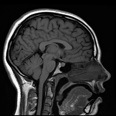
Magnetic Resonance Imaging (MRI) uses strong magnetic fields and radio waves accompanied by powerful computer technology to create a composite structural image of body tissue. Psychologists use them to learn about the structure and volume of tissue in the brain and spinal cord (the central nervous system (CNS). MRIs are safe aside from people with metal plates or screws inside their bodies (from surgery). In the past, risky, invasive surgery would have been necessary to determine issues with the CNS (though usually doctors were helpless and opted to do nothing but speculate) but now, techniques such as MRI provide a safe means of thoroughly examining brain structure.
While the way an MRI works is complex, it essentially relies on 2 powerful magnets that work together to align protons in water molecules (present in proteins, for example) so that they can then be measured by radio waves. These radio waves are detected and computed into numerous 2-D images that can be put together by a computer program to show cross-sections (slices) of the brain and even a 3-D representation.

<MORE>
MRI does not allow researchers to see brain functioning in real-time; they only allow the structure to be seen which also allows psychologists to compare the volume of certain areas and detect the size of lesions, for example. MRIs are useful for seeing structural change as a result of training or accidents, lesions, tumors, or other physiological abnormalities. Scans of American Footballers’ brains helped doctors see the long-term damage caused as a result of constantly receiving head trauma and concussions.
Since MRIs show structure, they are often used to compare two groups of people, such as Alzheimer’s or Parkinson’s sufferers with non-sufferers, to determine the main structures or areas affected. Since certain areas are associated with specific neurons that are activated by specific neurotransmitters it can provide important information about which neurotransmitters should be the focus of further research for potential treatment.

So, after aligning protons, the machine switches off the magnet and all protons return to normal. Therefore, the MRI does not cause any long-term effect on the brain or its functioning. For answering questions on imaging techniques in the biological unit you could use Maguire (2000), Draganski (2004), Corkin et al. (1997), for example. Other potential imaging techniques are the fMRI or PET scan, among others.
<LESS>